Intraoral Scanning Advances Every Dentist Should Know in 2025: Full-Arch Implant Restoration Insights for Dental Labs
In 2025, successful full-arch implant restorations depend on seamless collaboration between clinicians and dental labs. Labs translate digital impressions into physical solutions with accuracy and reliability, ensuring every step—from the initial scan to prosthesis delivery—produces consistent and high-quality outcomes.
Advances in Optical Accuracy and 3D Data Density for Implant Prostheses
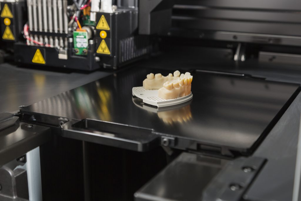
Today’s intraoral scanners provide ultra-high optical resolution and vastly improved 3D data density. These scanners capture even the smallest anatomical details, mapping teeth, implants, and soft tissues with unparalleled accuracy. Labs benefit by reducing the margin for error and creating prosthetics that fit more precisely with minimal adjustments.
State-of-the-art scanners now use advanced algorithms for stitching scan segments, improving data reliability—particularly valuable for full-arch cases, where even a small error can be magnified across multiple implants.
Scan Body Alignment and Library Validation for Multi-Implant Cases
The foundation of multi-implant success is correct scan body alignment. A single deviation can cause significant cumulative error in the final prosthesis. Labs now use standardized scan body libraries and AI-powered dental scanning tools to cross-check alignment, leveraging digital libraries validated for both shape and material.
Labs request dentists to use approved scan bodies and supply verification photos whenever possible to ensure the scan aligns with manufacturer specifications and library data.
Managing Full-Arch Accuracy: Reducing Cumulative Error Through Lab Protocols
A challenge unique to full-arch work is cumulative error—minor scanning inaccuracies that add up when fabricating large restorations. Leading labs address this with rigorous double scanning, overlay comparisons, and digital quality assurance checks. In practice, labs often build a “virtual case history,” comparing data points between scans, bite registrations, and facial scans to triangulate fit and function.
Soft-Tissue and Emergence Profile Capture for Optimal Marginal Fit
Modern scanners now effectively capture soft-tissue contours, emergence profiles, and even tissue dynamics. This is essential for lab technicians designing prosthetics that not only fit the implants but also blend naturally with the patient’s soft tissues. Precision in this area leads to restorations with optimal marginal fit, reducing tissue irritation and improving oral hygiene outcomes.
Color, Surface Texture, and Material Mapping for Esthetic and Functional Restorations
Intraoral imaging now extends beyond shape, providing comprehensive surface texture and color maps. Integrating this data enables labs to match neighboring teeth in shade and gloss, producing restorations that are both esthetic and functionally robust.
Patients benefit from teeth that look and feel natural, enhancing satisfaction and supporting long-term clinical success.
Scan Protocol Optimization: Techniques to Minimize Lab Adjustments and Remakes
Optimized scanning protocols are essential to minimize lab adjustments and avoid costly remakes. Labs advise clinicians to maintain a dry field, follow recommended scan path patterns, and validate scan completeness before sending data. These steps help labs begin fabrication with confidence, shortening turnaround times and reducing the number of clinical visits.
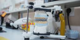
Verification Jigs: Translating Scan Data into Passive-Fit Frameworks
Verification jigs have become standard tools for translating digital impressions into physical models. Labs use them to confirm the passive fit of frameworks before final assembly. By constructing a verification jig from digital scan data, labs can test restorations for accurate, stress-free seating across all implants.
Integration with CAD/CAM and Milling or 3D Printing Workflows for Lab Consistency
Effective lab workflows now require compatibility with multiple digital design platforms. Labs routinely convert intraoral scan files directly into CAD/CAM or 3D-printing systems, streamlining production and ensuring high reproducibility.
Dental labs should choose scanner models and design software with open-architecture formats to maintain flexibility and adapt to evolving technologies.
Quality Control and Lab-Based Validation to Ensure Predictable Full-Arch Outcomes
Quality control is the backbone of modern dental lab success. Labs establish checklists for each client, verify scan data at every step, and maintain detailed logs of all cases. Before final assembly, lab-based validation ensures the restoration meets fit, function, and esthetic standards—delivering predictable, patient-approved resu0lts.
Conclusion
Advances in intraoral scanning, optimized scan protocols, and lab-driven validation provide clinicians and dental labs with powerful tools for excellence in 2025. Ongoing innovation, thorough quality control, and clear communication between team members will determine which practices excel in full-arch implant restorations. By embracing these trends, dental professionals ensure the best possible outcomes for every patient’s oral health.
Latest News
ICAM Photogrammetry: How Confident Dental Lab Delivers Once‑Impossible Full‑Arch Implant Precision
Advances in intraoral scanning, optimized scan protocols, and lab-driven validation provide clinicians and dental labs with powerful tools for excellence in 2025.
Zirconia Crowns: Benefits, Types, and Why Dentists Prefer Them
Zirconia crowns consist of zirconium dioxide, a crystalline ceramic engineered for exceptional toughness in restorative dentistry.
How a High-Quality Dental Lab Reduces Chair Time for Dentists
High-quality labs provide restorations that fit right away, so dentists can increase their weekly appointments by 15-25%. With digital tools and automation, workflows can move up…
Occlusal Scheme Design for Full-Arch Restorations: Balancing Function and Esthetics
Labs must often navigate the choice between functional occlusion, which focuses on ideal load distribution, longevity, and minimal stress on components, and esthetic occlusion.